
Upper Limb Trauma Surgery
Upper limb trauma surgery addresses fractures and injuries of the shoulder, arm, elbow, forearm, wrist, and hand using advanced fixation techniques. Dr. Sunny Dole specializes in precise, minimally invasive procedures to restore function and promote quick recovery.
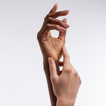
Evolution of Human Upper Limb and Hand Function
The evolution of the upper limb, particularly the human hand, is central to the development of human intelligence, creativity, and survival skills. In early primates, forelimbs were primarily used for climbing and locomotion, but as humans evolved into bipedal beings, the upper limbs were freed for more complex functions. This allowed the shoulder to gain a wider range of motion, the elbow to enable powerful flexion and extension, and the wrist to rotate and stabilize fine movements.
The human hand, with its opposable thumb and precisely aligned fingers, became a powerful tool capable of both strength and delicacy. This unique structure made it possible to create and use tools, develop language through gesture, build shelters, and perform skilled tasks like painting, surgery, or playing musical instruments. The dense sensory network in the fingertips also enhanced the brain's ability to perceive and interact with the environment.
Functionally, the upper limb is not just an anatomical feature but an essential system that integrates musculoskeletal strength, neuromuscular coordination, and sensory feedback—allowing humans to shape, manipulate, and understand the world around them in ways no other species can. This incredible evolution highlights why upper limb trauma care requires both anatomical understanding and surgical precision to restore not just movement but also quality of life.
Dr Sunny Dole has keen interest in Biomechanics and development of Upper Limb and Hand Function.
Managing Injuries from Shoulder to Finger : Dr Sunny Dole - Orthopedic Trauma Surgeon
Upper limb trauma encompasses a wide range of injuries involving the bones, joints, and soft tissues extending from the shoulder down to the fingertips. Each segment of the upper limb plays a vital role in motion, coordination, and function, and trauma to any of these areas can significantly impair daily activities and quality of life. Surgical intervention, when required, focuses on restoring alignment, stability, and mobility while minimizing long-term complications.
Shoulder Injuries often involve the clavicle (collarbone), scapula (shoulder blade), or proximal humerus (upper arm bone). Clavicle fractures are common in falls or road traffic accidents and may require plating for displaced fractures. Proximal humerus fractures—especially in older adults—may need surgical fixation with locking plates or even shoulder replacement in complex cases.
Humeral Shaft Fractures can occur from direct blows or twisting injuries. These fractures may be treated with intramedullary nailing or plating depending on the location and displacement. Special attention is given to protecting the radial nerve, which runs near the humeral shaft.
Elbow Injuries include distal humerus fractures, radial head fractures, and olecranon (elbow tip) fractures. The elbow is a complex hinge joint, and restoring its alignment is critical to avoid stiffness. Surgical fixation aims to preserve motion while stabilizing the bones.
Forearm Fractures typically involve the radius and ulna, either singly or in combination. These bones act together for rotational movements of the forearm (pronation and supination), so anatomical reduction and rigid fixation using plates and screws are necessary to restore function and alignment.
Wrist Fractures, particularly distal radius fractures, are among the most common upper limb injuries. These often result from falls on an outstretched hand. Depending on displacement and comminution, treatment may range from casting to surgical fixation with volar locking plates.
Hand and Finger Injuries include metacarpal and phalangeal fractures. These require precise alignment to maintain grip strength and dexterity. Intra-articular fractures or those causing rotational deformities are usually treated surgically with pins, screws, or mini-plates.
From shoulder dislocations to complex hand injuries, upper limb trauma surgery demands a meticulous and individualized approach. The goal is not only to heal bones but also to preserve the intricate function of the arm and hand. Dr. Sunny Dole brings expertise in upper limb trauma surgery, ensuring timely and accurate treatment tailored to each patient’s injury and needs.
Wrist Fracture : Recent Advances in Treatment Options
Wrist fractures are common injuries, and recent advances in treatment options aim to improve healing, reduce complications, and restore function more effectively. These advances span both non-surgical and surgical approaches, with a growing emphasis on minimally invasive techniques and personalized treatment strategies.
Recent Advances in Diagnosis
-
Advanced Imaging: While X-rays remain the initial diagnostic tool, advanced imaging techniques like CT scans and MRI are increasingly used to provide more detailed information about the fracture pattern, associated soft tissue injuries (ligaments, tendons), and occult fractures not visible on plain radiographs. MRI is particularly useful for detecting early scaphoid fractures and ligamentous injuries.
-
Point-of-Care Ultrasound (POCUS): In pediatric distal radius fractures, POCUS has emerged as a safe and efficient means for diagnosis, potentially reducing the need for radiography and shortening emergency department stays. However, widespread adoption may be limited by the initial costs of training and equipment.
Recent Advances in Non-Surgical Treatment
-
Challenging Rigid Immobilization: For certain types of wrist fractures, particularly torus fractures in children, recent evidence challenges the traditional need for rigid immobilization. Studies have shown that simple bandages may be equivalent to casts or splints in terms of pain and functional outcomes. This approach aims to minimize stiffness and allow earlier return to function.
-
Optimized Casting Techniques: Research continues to refine casting techniques, although there is no clear consensus on the optimal wrist position for immobilization. Some advocate for supination to counter deforming forces, while others suggest pronation. Most surgeons use some degree of palmar flexion based on biomechanical principles.
Recent Advances in Surgical Treatment
-
Minimally Invasive Techniques:
-
Wrist Arthroscopy: This technique has seen significant advancements, allowing for the diagnosis and treatment of various wrist disorders through small incisions using a camera and specialized instruments. It is increasingly used for:
-
Fracture Reduction and Internal Fixation (ARIF): Arthroscopy can assist in the precise reduction (realignment) of fracture fragments and facilitate minimally invasive internal fixation with screws or pins.
-
Scaphoid Fracture Fixation: Arthroscopic techniques for scaphoid fracture fixation are evolving, offering potential benefits of reduced soft tissue disruption and improved visualization.
-
TFCC Repair: Arthroscopy is now a mainstay for treating triangular fibrocartilage complex (TFCC) tears, with outcomes comparable to open surgery.
-
Ligament Repair and Reconstruction: Chronic scapholunate ligament injuries, a challenging area, are seeing the development of anatomical front-and-back reconstruction techniques using grafts, with early promising results.
-
-
Intramedullary Nailing: For unstable extra-articular distal radius fractures, the Distal Radius Intramedullary Nail (DRIM-Nail) is a relatively new option. Inserted through a small incision, it aims to provide stable fixation while minimizing soft tissue irritation and allowing for early mobilization.
-
-
Internal Fixation with Plates and Screws: This remains a common and effective surgical option for many wrist fractures, particularly unstable, intra-articular, and comminuted fractures. Advances include:
-
Low-Profile Volar Plates: These plates are designed to minimize tendon irritation and rupture, a known complication of traditional volar plating.
-
Dorsal Plating: Improved implant designs now allow for safer dorsal fixation in cases with dorsal comminution.
-
-
External Fixation: External fixators, with pins placed through the skin into the bone and connected to an external frame, are used for complex fractures, open fractures, and situations where internal fixation is not feasible.
-
Bone Grafting and Biologics:
-
Bone Grafts: Autografts (from the patient's own body, such as the distal radius or iliac crest) and allografts (from a donor) are used to promote healing in non-unions or fractures with bone loss.
-
Vascularized Bone Grafts (VBGs): For scaphoid non-unions, VBGs, often based on the palmar carpal artery or pronator quadratus pedicle, have shown high union rates, particularly in challenging cases.
-
Orthobiologics: Emerging therapies like mesenchymal stem cells and bone morphogenetic proteins are being investigated for their potential to enhance healing in scaphoid non-unions and revision surgeries. Calcium phosphate bone substitutes are also used to fill defects and promote bone ingrowth.
-
Recent Advances in Scaphoid Fracture Treatment
-
Debate on Early Surgical Fixation: There is ongoing debate about the optimal management of non-displaced scaphoid waist fractures, comparing early surgical fixation with conservative treatment in a cast. Some studies suggest faster healing and return to work with surgery, while others show similar functional outcomes in the long term.
-
Importance of Screw Placement: For surgical fixation, precise screw placement in the central axis of the scaphoid is crucial for stability and healing. Both dorsal and volar approaches are used, with the volar approach often preferred for its exposure and lower risk to the vascular supply.
-
Management of Non-Union: Treatment options for scaphoid non-union include open reduction and internal fixation with bone grafting (iliac crest or distal radius), vascularized bone grafts, and salvage procedures like proximal row carpectomy or wrist fusion in cases with arthritis. Arthroscopic techniques for non-union repair are also showing promising results with less morbidity.
Rehabilitation
-
Early Mobilization: Following surgical fixation, early, controlled mobilization is increasingly emphasized to reduce stiffness and promote functional recovery.
-
Hand Therapy: While home exercise programs may be sufficient for simple distal radius fractures treated with casting, formal hand therapy is often recommended after surgery to improve range of motion, strength, and function. Studies have shown short-term benefits of therapy in improving outcomes.
In summary, recent advances in wrist fracture treatment focus on more accurate diagnosis, less invasive surgical techniques, personalized treatment approaches based on fracture characteristics and patient factors, and optimized rehabilitation protocols to achieve better long-term functional outcomes.
Overview of Brachial Plexus Injuries
Brachial plexus injury refers to damage to the complex network of nerves that originate from the spinal cord in the neck (C5–T1) and control muscle movements and sensation in the shoulder, arm, and hand. These injuries can result from trauma such as motor vehicle accidents, falls, or penetrating injuries, and can range from mild stretching (neurapraxia) to complete nerve rupture or avulsion from the spinal cord. Symptoms depend on the level and severity of the injury, including weakness, loss of sensation, or complete paralysis of the upper limb. Early diagnosis using clinical evaluation, electromyography (EMG), and imaging (MRI or CT myelography) is essential. Treatment varies from conservative therapy and physiotherapy in mild cases to surgical repair, nerve grafting, or nerve transfers in severe injuries.
Surgical Treatment of Elbow Fractures
Surgical treatment for elbow fractures is often necessary when the broken bones are displaced, unstable, or when there are open fractures (bone protruding through the skin). The primary goals of surgery are to realign the fractured bone fragments (reduction) and stabilize them in the correct position until they heal. This is typically achieved through Open Reduction and Internal Fixation (ORIF).
Here's a detailed overview of surgical treatment for elbow fractures:
1. Indications for Surgery:
-
Displaced fractures: When the broken bone fragments have shifted out of their normal alignment.
-
Unstable fractures: Fractures that are likely to move out of alignment even with casting.
-
Open fractures: These have a high risk of infection and require immediate surgical cleaning and stabilization.
-
Intra-articular fractures: Fractures that involve the joint surface, as precise alignment is crucial to prevent post-traumatic arthritis.
-
Fractures with associated injuries: Such as nerve or blood vessel damage that may require surgical exploration and repair.
-
Non-union: When a fracture fails to heal properly over time, surgery with bone grafting may be needed.
2. Surgical Procedure (Open Reduction and Internal Fixation - ORIF):
-
Anesthesia: The surgery is usually performed under general anesthesia, so the patient is asleep and pain-free. Regional anesthesia (nerve block) with or without sedation may also be used.
-
Surgical Incision: The surgeon will make an incision over the fractured area. The location and size of the incision depend on the type and location of the fracture. Careful attention is paid to avoid damaging surrounding nerves, blood vessels, and soft tissues.
-
Fracture Reduction: The surgeon will carefully manipulate and reposition the broken bone fragments back into their normal anatomical alignment. This is the "reduction" part of the procedure.
-
Internal Fixation: Once the bones are realigned, they need to be held in place to allow for proper healing. This is achieved using various internal fixation devices:
-
Plates and Screws: Metal plates are contoured to fit the shape of the bone and are secured to the bone fragments with screws. This provides strong and stable fixation, especially for complex or comminuted (multiple fragments) fractures. Low-profile plates are often used to minimize irritation to surrounding soft tissues.
-
Screws: Lag screws can be used to compress fracture fragments together, promoting healing, particularly in certain types of fractures like some radial head fractures.
-
Wires (K-wires): Kirschner wires are thin metal wires that can be used to temporarily or permanently hold small bone fragments in place. They are often used in conjunction with other fixation methods. Tension band wiring is a specific technique using wires to compress olecranon fractures.
-
Intramedullary Nailing: While less common in elbow fractures compared to long bone fractures, intramedullary nails (rods inserted into the hollow center of the bone) can be used for certain shaft fractures of the humerus near the elbow.
-
External Fixation: In cases of open fractures, severe soft tissue injury, or highly comminuted unstable fractures where internal fixation might be risky or not immediately feasible, an external fixator may be used. This involves placing pins through the skin into the bone fragments and connecting them to an external frame to stabilize the elbow. External fixators can be temporary or definitive.
-
-
Other Repairs: If there are associated injuries, such as ligament or tendon tears, these may also be repaired during the surgery.
-
Wound Closure: After the fracture is fixed and any other necessary repairs are made, the surgeon will close the layers of skin and muscle with sutures or staples. A sterile dressing will be applied. In some cases, a drain may be placed to prevent fluid buildup.
-
Splint or Brace: Following surgery, the elbow is usually immobilized in a splint or brace to protect the repair and allow for initial healing.
3. Specific Types of Elbow Fractures and Surgical Approaches:
-
Olecranon Fractures (Tip of the elbow): Often require ORIF with tension band wiring or plate and screws to restore the extensor mechanism of the elbow.
-
Radial Head Fractures (Part of the forearm bone at the elbow): Treatment depends on the number of fragments and displacement. Non-displaced fractures may be treated non-surgically. Displaced fractures may require ORIF with small plates and screws or, in severely comminuted cases, radial head excision or replacement with a prosthesis.
-
Distal Humerus Fractures (Lower end of the upper arm bone): These are complex fractures often requiring ORIF with plates and screws on both the medial and lateral columns of the distal humerus to reconstruct the joint surface and provide stability.
-
Coronoid Fractures (Projection on the ulna): These fractures are important for elbow stability and often require ORIF with screws or suture anchors, especially in the context of elbow dislocations.
4. Post-operative Care and Rehabilitation:
-
Immobilization: The duration of immobilization in a splint or brace varies depending on the fracture pattern and the type of fixation used.
-
Pain Management: Pain medication will be prescribed to manage post-operative pain.
-
Swelling Control: Elevating the arm and applying ice can help reduce swelling.
-
Early Mobilization: Once the initial healing phase allows, early and controlled range of motion exercises are crucial to prevent elbow stiffness, a common complication after elbow fractures. This is often guided by a physical therapist.
-
Physical Therapy: A structured physical therapy program is essential to regain full range of motion, strength, and function of the elbow and arm. This may involve various exercises, manual therapy techniques, and modalities.
-
Weight-bearing Restrictions: There will be restrictions on lifting and using the operated arm for a period of time.
-
Follow-up Appointments: Regular follow-up appointments with the surgeon are necessary to monitor healing and progress.
-
Implant Removal: In some cases, if the hardware causes irritation or pain after the fracture has healed, it may be surgically removed, but this is not always necessary.
5. Potential Risks and Complications of Surgery:
-
Infection: As with any surgery, there is a risk of infection at the surgical site.
-
Nerve and Blood Vessel Damage: There is a small risk of injury to the nerves and blood vessels around the elbow during surgery. Temporary numbness or weakness can occur.
-
Stiffness: Elbow stiffness is a common complication after elbow fractures, even with surgery and rehabilitation.
-
Non-union or Malunion: The fracture may fail to heal properly (non-union) or heal in a poor position (malunion), potentially requiring further surgery.
-
Hardware Failure: The plates, screws, or wires may break or shift.
-
Post-traumatic Arthritis: Damage to the joint cartilage at the time of the fracture can lead to arthritis over time.
-
Heterotopic Ossification: Bone may form in the soft tissues around the elbow, restricting movement.
-
Pain at the Hardware Site: The internal fixation devices may sometimes cause discomfort.
-
Complex Regional Pain Syndrome (CRPS): A rare condition characterized by chronic pain, swelling, and changes in skin color and temperature.
The decision to proceed with surgical treatment for an elbow fracture is made by an orthopedic surgeon based on a thorough evaluation of the fracture, the patient's overall health, and their functional demands. The specific surgical technique and post-operative rehabilitation plan are tailored to the individual patient and the nature of their injury to optimize healing and functional recovery.

Shoulder Fracture Surgery : What are newer treatment options?
Shoulder fractures—comprising fractures of the clavicle, scapula, and proximal humerus—are common injuries, especially among the elderly due to falls and in young adults due to high-energy trauma. The shoulder is a highly mobile joint with complex anatomy, and restoring its function after a fracture requires a deep understanding of bone healing principles, biomechanics, and soft tissue preservation.
Types of Shoulder Fractures
-
Clavicle Fracture – Often occurs in the midshaft and can usually be treated conservatively, although displaced or comminuted fractures may require surgical fixation.
-
Proximal Humerus Fracture – Common in osteoporotic patients, it may involve one to four parts as per the Neer classification. Management depends on displacement and vascularity of the humeral head.
-
Scapular Fracture – Rare, usually associated with high-energy trauma, and often requires CT for diagnosis. Most can be managed non-operatively unless displaced.
Treatment Principles
The core principles of managing shoulder fractures include:
-
Anatomical reduction of the fracture
-
Stable fixation while preserving soft tissue
-
Early mobilization to prevent stiffness
-
Restoration of joint congruity, especially in articular fractures
Surgical intervention is often required for displaced fractures or when conservative treatment fails. Techniques include open reduction and internal fixation (ORIF), minimally invasive surgery, and in selected cases, shoulder arthroplasty (replacement).
Recent Advances in Treatment & Implant Design
-
Locking Plate Technology
The use of locking compression plates (LCPs) has revolutionized shoulder fracture fixation, especially for osteoporotic bone. Locking screws provide angular stability, reducing the risk of implant failure and loss of fixation. -
Intramedullary Nails for Proximal Humerus
Modern nails allow stable fixation with minimal soft tissue disruption. Some systems come with variable angle locking and suture holes for rotator cuff integration. -
Augmented Fixation Techniques
For comminuted or osteoporotic fractures, the use of bone grafts or cement augmentation improves the biomechanical strength of fixation. -
Reverse Shoulder Arthroplasty (RSA)
In elderly patients with complex, non-reconstructible fractures of the proximal humerus, RSA provides better outcomes than conventional hemiarthroplasty, especially when the rotator cuff is deficient. -
Minimally Invasive Approaches
The deltoid-sparing or deltopectoral approach minimizes soft tissue injury, reduces blood loss, and improves recovery times.
Rehabilitation and Recovery
Rehabilitation is a critical component of shoulder fracture management. Early passive motion is usually initiated within the first 1–2 weeks after surgery, followed by active-assisted and active range of motion over the next few weeks. Strengthening exercises begin once fracture union is evident, typically around 6–8 weeks post-surgery.
-
Timeline Summary:
-
0–2 weeks: Sling immobilization, passive range of motion
-
2–6 weeks: Active-assisted exercises
-
6–12 weeks: Active exercises and strengthening
-
Full functional recovery: 3–6 months (may vary with fracture type and patient age)
-
Why Choose Dr. Sunny Dole for Shoulder Fracture Treatment?
Dr. Sunny Dole is an experienced orthopedic trauma and joint reconstruction surgeon in Pune who offers cutting-edge treatment options for shoulder fractures. He uses advanced implants and minimally invasive techniques tailored to the patient’s age, bone quality, and activity level. With a strong focus on early rehabilitation and long-term function, patients under Dr. Dole’s care experience faster recovery and reduced complications.
Precision in Motion: Expert Care for Hand Trauma Surgery
The human hand is a marvel of intricate anatomy and biomechanics, responsible for tasks that range from delicate fine motor movements to powerful gripping functions. When hand trauma occurs—due to accidents, falls, machinery injuries, or sports—it can severely impact quality of life and productivity. At the forefront of managing such complex injuries is Hand Trauma Surgery, a specialized field combining orthopedic precision, microsurgical expertise, and modern rehabilitation principles.
Types of Hand Trauma
Hand injuries can involve bones, tendons, ligaments, nerves, blood vessels, and skin. Common hand trauma conditions include:
-
Metacarpal and Phalangeal Fractures: Fractures of the hand bones often result from direct trauma or falls. Treatment may include splinting or surgical fixation using mini-plates, screws, or K-wires to ensure proper alignment and quick recovery.
-
Tendon Injuries: Lacerations or ruptures of flexor and extensor tendons affect the ability to bend or straighten fingers. Surgical repair requires precise handling and rehabilitation to restore function.
-
Nerve and Vessel Injuries: Trauma involving digital nerves or arteries can lead to numbness, pain, or loss of circulation. Microsurgical techniques are used for delicate repair under magnification.
-
Crush Injuries and Amputations: Severe trauma from industrial or road traffic accidents may cause partial or total hand amputations. Early debridement, soft tissue coverage, and, in some cases, replantation are essential.
-
Infection and Open Injuries: Contaminated wounds can quickly lead to complications if not treated with proper debridement and antibiotics.
Advances in Hand Trauma Surgery
Recent innovations in hand trauma care have significantly improved outcomes:
-
Low-Profile Implant Designs: Modern implants for small bones are anatomically contoured and made of bio-compatible materials, allowing stable fixation without bulk.
-
Microsurgical Techniques: Use of microscopes and fine sutures enables repair of tiny nerves and vessels, critical for sensory and functional restoration.
-
Minimally Invasive Procedures: In select cases, arthroscopic or percutaneous techniques reduce tissue trauma and enhance recovery.
-
Biological Adjuncts: Use of Platelet-Rich Plasma (PRP) and bone graft substitutes accelerates healing and reduces post-operative stiffness.
Principles of Hand Trauma Management
Successful hand trauma care follows key surgical principles:
-
Timely Intervention: Early diagnosis and intervention prevent complications like joint stiffness and tendon adhesions.
-
Anatomical Realignment: Bone fractures must be aligned with great precision to preserve joint congruity and motion.
-
Stable Fixation with Early Mobilization: Using implants that allow immediate motion prevents stiffness and improves outcomes.
-
Preservation of Soft Tissue and Blood Supply: Essential for healing, especially in crush or degloving injuries.
-
Multidisciplinary Rehabilitation: Post-operative physiotherapy plays a vital role in regaining hand strength, dexterity, and function.
Recovery and Rehabilitation
Rehabilitation begins immediately after surgery and is tailored to the injury type. Splinting, guided exercises, and functional retraining ensure that patients regain maximal independence and hand use. The recovery timeline varies—minor fractures may heal in 4–6 weeks, while complex reconstructions may require several months of therapy.
Conclusion
Whether you're dealing with a simple finger fracture or a complex hand crush injury, precision in motion begins with expert surgical care. Dr. Sunny Dole offers advanced hand trauma surgery in Pune with a focus on evidence-based techniques, microsurgical excellence, and individualized rehabilitation plans. Trust your hands to hands that heal—with precision, compassion, and care.